Shop
No results found
You can try clearing any filters or head to our store's home
No results found
You can try clearing any filters or head to our store's home
Endocrinology is the branch of clinical medicine and laboratory science focused on the endocrine system—the network of glands that produce and secrete hormones into the bloodstream.
These chemical messengers regulate vital physiological processes, including metabolism, growth, tissue function, reproduction, sleep, and mood. In diagnostic laboratories, endocrinology panels utilize high-sensitivity immunoassay and mass spectrometry techniques to detect hormonal imbalances and glandular disorders.
Thyroid & Parathyroid: Produces T3, T4, and TSH to regulate metabolic rate, energy expenditure, and calcium homeostasis via PTH (Parathyroid Hormone).
Pancreas (Endocrine Function): Secretes Insulin and Glucagon to maintain tight glycemic control and cellular glucose uptake.
Adrenal Glands: Synthesizes Cortisol (stress response and metabolism), Aldosterone (blood pressure and electrolyte balance), and Adrenaline.
Pituitary & Hypothalamus: The “master regulators” that release tropic hormones (ACTH, TSH, LH, FSH, GH, Prolactin) to command peripheral endocrine organs.
Reproductive Axis (Gonads): Produces Testosterone, Estrogen, and Progesterone driving sexual development, fertility, and secondary tissue maintenance.
Occupational endocrinology focuses on how workplace exposures, shift work, and chronic physical stressors disrupt hormonal synthesis and metabolic regulation:
| Workplace Factor | Affected Endocrine Axis | Clinical & Diagnostic Consequence |
| Endocrine Disrupting Chemicals (EDCs) | Thyroid & Reproductive Hormones | Pesticides, phthalates, and bisphenols interfering with hormone receptor binding |
| Night Shift & Rotational Work | Melatonin & HPA Axis (Cortisol) | Circadian rhythm disruption leading to insulin resistance and metabolic syndrome |
| Heavy Metal Toxicants (Lead, Cadmium) | Pituitary-Gonadal & Adrenal Axis | Impairment of fertility parameters and altered stress-response mechanisms |
| Extreme Heat / Physical Stress | Vasopressin (ADH) & Aldosterone | Fluid retention imbalance, chronic adrenal fatigue, and electrolyte dysfunction |
Early Metabolic Detection: Identifies subclinical hormone imbalances (such as early insulin resistance or subclinical hypothyroidism) long before physical organ damage occurs.
High Precision Immunoassays: Utilizes ECLIA (Electrochemiluminescence) and LC-MS/MS (Liquid Chromatography-Mass Spectrometry) for pinpoint accuracy at low hormonal concentrations.
Comprehensive Health Surveillance: Essential for executive physicals, fertility assessments, and long-term toxicity monitoring in industrial work environments.
Biochemistry (or Clinical Chemistry) is the branch of medical laboratory science that analyzes chemical reactions, metabolic pathways, and biological fluids to evaluate organ function and metabolic health.
By measuring enzymes, hormones, electrolytes, proteins, and metabolic waste products in serum or plasma, biochemistry provides essential quantitative data for routine health assessments, acute disease diagnosis, and chronic condition monitoring.
Liver Function Tests (LFTs): Evaluates hepatic integrity and biliary excretion using markers like ALT, AST, Alkaline Phosphatase (ALP), Bilirubin, and Albumin.
Renal Function Tests (RFTs / Kidney Panel): Measures kidney filtration capacity and fluid balance using Blood Urea Nitrogen (BUN), Serum Creatinine, Uric Acid, and Electrolytes (Na+, K+, Cl-).
Lipid Profile: Assesses cardiovascular risk factors by quantifying Total Cholesterol, Triglycerides, HDL (high-density lipoprotein), and LDL (low-density lipoprotein).
Endocrine & Diabetes Diagnostics: Monitors metabolic regulation through Fasting Blood Glucose, HbA1c (glycated hemoglobin), Insulin, and Thyroid function markers (TSH, Free T3, Free T4).
In workplace health settings, clinical biochemistry acts as an early warning system to detect organ strain or metabolic disruption caused by physical and chemical occupational hazards:
| Occupational Hazard | Biochemistry Test Panel | Clinical / Preventative Objective |
| Industrial Solvents & Chemicals | Liver Function & Enzyme Panels | Early detection of chemical-induced hepatotoxicity in factory workers |
| Heavy Metal Exposure (Lead, Cadmium) | Renal Biomarkers & Serum Electrolytes | Screening for early tubular damage or nephrotoxicity |
| Organophosphate Pesticides | Serum / Red Blood Cell Cholinesterase | Assessing enzyme inhibition in agricultural and chemical handlers |
| Heat Stress & Strenuous Physical Work | Electrolyte Panel & Serum Creatine Kinase (CK) | Monitoring dehydration, rhabdomyolysis risk, and muscle tissue breakdown |
High Automation & Throughput: Automated clinical chemistry analyzers process high sample volumes quickly, making them ideal for large-scale employee wellness screenings.
Quantitative Baseline Metrics: Establishes objective numerical baselines for workers to track subtle organ function shifts over time.
Broad Screening Capability: A single blood sample yields comprehensive insights into multiple physiological systems simultaneously.
Phlebotomy is the clinical practice of puncturing a vein (venipuncture) to collect blood samples for diagnostic testing, transfusions, research, or blood donations.
As the primary entry point for laboratory medicine, phlebotomy is critical for pre-analytical quality control—ensuring specimen integrity, proper labeling, and accurate test results across all diagnostic departments.
Venipuncture: The most common technique, typically drawing blood from the antecubital fossa (inner elbow) using a evacuated tube system (Vacutainer) or a butterfly needle assembly.
Capillary Collection (Dermal Puncture): Obtaining small capillary blood volumes via heel sticks (infants) or finger pricks (point-of-care testing, blood glucose, hemoglobin).
Arterial Blood Draw: Performed by specialized clinicians (usually from the radial artery) specifically to measure arterial blood gases (ABGs) for respiratory and acid-base evaluation.
Phlebotomy relies on standardized, color-coded collection tubes containing specific additives, anticoagulants, or clot activators:
| Tube Top Color | Additive / Coating | Primary Diagnostic Tests |
| Lavender / Purple | EDTA (Anticoagulant) | Complete Blood Count (CBC), HbA1c, Blood Smear |
| Red / Gold (SST) | Clot Activator & Gel Separator | Routine Chemistry, Lipid Panel, Thyroid, Serology |
| Light Blue | Sodium Citrate | Coagulation studies (PT/INR, PTT, D-Dimer) |
| Green | Sodium / Lithium Heparin | STAT Plasma Chemistry, Electrolytes, Ammonia |
| Gray | Sodium Fluoride & Potassium Oxalate | Glucose tolerance testing, Blood Alcohol Levels |
In workplace health settings, efficient phlebotomy operations support both routine employee wellness and specialized safety compliance:
On-Site Health Screenings: Rapid collection setups for high-volume annual employee physicals, executive health checks, and baseline lab panels.
Biological Hazard Monitoring: Collecting samples to measure heavy metal exposures (e.g., lead, cadmium) and chemical toxin levels in industrial workers.
Pre-Employment & Drug Screening: Chain-of-custody collection procedures for occupational drug panels and post-accident toxicological investigations.
Molecular Biology and Genetics is the field of life science focused on understanding the structure, function, and manipulation of nucleic acids (DNA and RNA) and proteins. While genetics examines heredity, trait inheritance, and gene variations, molecular biology investigates the physical and chemical mechanisms that govern gene expression, DNA replication, and cellular function at a molecular level.
Polymerase Chain Reaction (PCR & RT-qPCR): Amplifying specific target DNA sequences to detectable levels, enabling precise pathogen identification and gene expression analysis.
Next-Generation Sequencing (NGS): High-throughput sequencing of entire genomes or targeted gene panels to detect inherited mutations, somatic variations, and structural rearrangements.
Recombinant DNA & Gene Editing: Techniques such as molecular cloning and CRISPR-Cas9 to modify genetic sequences for therapy, biomanufacturing, and research.
Gene Expression Profiling: Quantifying RNA transcripts to understand cellular responses to external stressors, biological toxins, or disease progression.
In modern diagnostic laboratories and labor medicine, molecular genetics provides high-sensitivity, high-specificity testing for early intervention and risk assessment:
| Application | Molecular Testing Method | Clinical / Occupational Utility |
| Occupational Toxicology | Genotoxicity & DNA Adduct Profiling | Screening workers for molecular damage caused by carcinogens, radiation, or industrial solvents |
| Infectious Disease Surveillance | Multiplex Real-Time PCR | Rapid detection of respiratory pathogens, bloodborne viruses, and occupational biological hazards |
| Somatic Cancer Screening | Liquid Biopsy & Biomarker Sequencing | Detecting circulating tumor DNA (ctDNA) for early oncological detection and targeted therapy matching |
| Pharmacogenomics (PGx) | Targeted Genotyping Panels | Analyzing genetic variants in drug-metabolizing enzymes (e.g., CYP450) to optimize medication dosing |
Unmatched Sensitivity: Can detect tiny trace amounts of viral, bacterial, or mutated genetic material long before physical symptoms appear.
High Specificity: Targets unique genomic sequences, drastically reducing false positives and cross-reactivity.
Precision Medicine Integration: Enables targeted therapies based on an individual’s unique genetic profile or disease driver mutations.
Cytology (or Cytopathology) is the branch of pathology that studies individual cells or small clusters of cells to diagnose diseases, most notably precancerous conditions, cancers, infections, and inflammatory states.
Unlike histology, which examines intact tissue architecture obtained via biopsy, cytology analyzes loose cells collected from bodily fluids, mucosal scrapings, or fine-needle aspirations.
Exfoliative Cytology: Examination of cells that naturally shed or are gently scraped/brushed from epithelial surfaces.
Gynecologic: Pap smears for cervical cancer screening and HPV detecting.
Non-Gynecologic: Sputum samples, urine cytology, cerebrospinal fluid (CSF), pleural/peritoneal fluid, and bronchial washings.
Aspiration Cytology (Fine-Needle Aspiration – FNA): Using a thin needle attached to a syringe to extract fluid or tissue fragments directly from solid masses or nodules (e.g., thyroid nodules, breast lumps, lymph nodes, salivary glands).
In occupational health settings, cytology serves as an essential screening tool for early detection of carcinogen-induced cellular changes in workers exposed to industrial hazards:
| Exposure Risk | Cytological Screening Test | Diagnostic Objective |
| Aromatic Amines / Dyes / Solvents | Urine Cytology | Screening chemical plant workers for urothelial dysplasia or bladder carcinoma |
| Asbestos / Silica / Industrial Dusts | Sputum Cytology & Bronchoalveolar Lavage (BAL) | Detecting premalignant changes in bronchial epithelium or identifying ferruginous bodies |
| Heavy Metals & Toxic Fumes | Nasal / Mucosal Brushing | Assessing chronic inflammation, squamous metaplasia, and upper respiratory tract injury |
Minimally Invasive: Samples are collected with minimal discomfort compared to surgical tissue biopsies.
Fast Turnaround: Staining procedures (such as Papanicolaou or Giemsa stains) can yield diagnostic results rapidly.
Early Detection: Identifies cellular atypia and dysplastic changes before tumors present symptoms or visible mass lesions.
| Area | Primary Conditions & Functions | Common Tests |
| Red Blood Cell Disorders | Anemia (iron-deficiency, sickle cell, aplastic), Thalassemia, Polycythemia | Full Blood Count (FBC/CBC), Ferritin, Reticulocyte count |
| Coagulation & Hemostasis | Hemophilia, Von Willebrand disease, Deep Vein Thrombosis (DVT) | Prothrombin Time (PT/INR), APTT, D-Dimer |
| Haematological Malignancies | Leukemia, Lymphoma (Hodgkin & Non-Hodgkin), Multiple Myeloma | Bone marrow biopsy, Flow cytometry, Cytogenetics |
| Transfusion Medicine | ABO & Rh blood typing, crossmatching, blood product preparation | Blood grouping, Antibody screening |
Parasitology is the branch of biology and medicine dedicated to studying parasites, their host organisms, and the ecological and biological relationships between them.
Unlike mutualistic or commensal relationships, a parasite lives in or on a host organism, obtaining nourishment at the host’s expense.
In medical and biological sciences, parasitology is generally categorized into three main areas based on the organism type:
Studies single-celled eukaryotic organisms. These often reproduce rapidly within a host and can cause severe acute infections.
Intestinal Protozoa: Entamoeba histolytica (amoebic dysentery), Giardia lamblia (giardiasis), Cryptosporidium.
Blood & Tissue Protozoa: Plasmodium species (malaria), Trypanosoma (sleeping sickness & Chagas disease), Leishmania, Toxoplasma gondii.
Studies multicellular, macroscopic worms. Unlike protozoa, adult helminths generally do not multiply within the human body.
Nematodes (Roundworms): Ascaris lumbricoides, hookworms (Necator americanus), pinworms (Enterobius vermicularis), and filarial worms.
Cestodes (Tapeworms): Taenia saginata (beef tapeworm), Taenia solium (pork tapeworm), Echinococcus.
Trematodes (Flukes): Schistosoma (blood flukes causing schistosomiasis/bilharzia), Fasciola hepatica (liver fluke).
Focuses on organisms that live on the outer surface of the host or act as vectors transmitting pathogens.
External Parasites: Lice (Pediculus humanus), scabies mites (Sarcoptes scabiei), bedbugs, and ticks.
Vectors: Anopheles mosquitoes (malaria vector), tsetse flies (sleeping sickness vector), and triatomine bugs.
| Concept | Description | Example |
| Definitive Host | The host in which the parasite reaches sexual maturity and reproduces sexually. | Anopheles mosquito for Plasmodium; Humans for Taenia solium. |
| Intermediate Host | A host required for parasite development, where asexual reproduction or larval stages occur. | Humans for Plasmodium; Pigs for Taenia solium. |
| Reservoir Host | An animal species that harbors a pathogen and serves as a source of infection for humans. | Snails for Schistosoma; Dogs for Leishmania. |
| Vector | An organism (typically an arthropod) that transmits a parasite from one host to another. | Ticks transmitting Babesia. |
Microscopy: Stool examinations (O&P – Ova and Parasites), thick and thin blood smears (for malaria/filaria).
Serology & Rapid Tests: Antigen/antibody detection (e.g., Malaria RDTs, ELISA).
Molecular Diagnostics: PCR testing for species identification and drug-resistance markers.
Histology (also known as microscopic anatomy) is the branch of biology and medicine that studies the microscopic structure of biological tissues, revealing how individual cells and extracellular matrix components assemble to form functional organs.
All human organs are composed of four basic functional tissue categories:
Epithelial Tissue: Covers body surfaces, lines internal cavities and organs, and forms glands. It serves protective, absorptive, and secretory functions (e.g., skin epidermis, intestinal lining).
Connective Tissue: Supports, binds, and protects other tissues and organs. It consists of cells suspended in an extracellular matrix and includes loose connective tissue, adipose (fat), cartilage, bone, and blood.
Muscle Tissue: Formed by specialized contractile cells capable of generating mechanical force. Divided into three types:
Skeletal muscle: Voluntary and striated (attached to bones).
Cardiac muscle: Involuntary and striated (found in the heart wall).
Smooth muscle: Involuntary and non-striated (lines blood vessels and digestive tracts).
Nervous Tissue: Composed of neurons (which transmit electrical impulses) and glial cells (which support and insulate neurons). Makes up the brain, spinal cord, and peripheral nerves.
To view tissues under an optical or electron microscope, specimens undergo a multi-step preparation process:
Fixation: Uses chemicals (commonly formaldehyde) to preserve cell structure and prevent decay.
Embedding & Sectioning: Tissue is dehydrated, embedded in paraffin wax, and sliced into ultra-thin sections (typically 4–5 micrometers thick) using a microtome.
Staining: Raw tissue slices are nearly transparent. Stains provide contrast:
H&E (Hematoxylin and Eosin): The standard gold-standard stain. Hematoxylin stains cell nuclei blue/purple, while Eosin stains cytoplasm and extracellular proteins pink.
Special Stains: Such as Masson’s trichrome (highlights collagen) or Periodic acid–Schiff (PAS, highlights carbohydrates and mucins).
Microbiology is the scientific study of microscopic organisms—those that are unicellular, multicellular, or acellular. It encompasses the structure, function, ecology, classification, and clinical relevance of bacteria, viruses, fungi, protozoa, and algae.
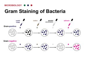
Microbiology is divided into sub-disciplines based on the specific group of organisms studied:
Bacteriology: The study of bacteria, their cell wall composition, metabolic pathways, and role in health and disease.
Virology: The study of sub-microscopic genetic elements (DNA or RNA) enclosed in protein coats (viruses) and sub-viral agents.
Mycology: The study of fungi, including yeasts, molds, and mushrooms, focusing on fungal pathogenesis, mycotoxins, and industrial fermentation.
Parasitology: Focuses on pathogenic protozoa and helminths (parasitic worms), their life cycles, vectors, and host interactions.
Immunology: Often grouped with medical microbiology, studying how host organisms defend against pathogen invasion.
In medical settings, identifying pathogens involves systematic diagnostic workflows to guide antimicrobial treatment:
| Technique | Method / Purpose | Common Examples |
| Microscopy & Staining | Direct visual examination and cellular wall differentiation | Gram stain, Acid-Fast (Ziehl-Neelsen) stain, KOH wet mount |
| Culture & Isolation | Growing organisms on selective/differential media to observe colony morphology | Blood agar, MacConkey agar, Sabouraud dextrose agar |
| Biochemical Testing | Evaluating metabolic enzymes and nutrient utilization | Catalase, Coagulase, Oxidase, Indole, Triple Sugar Iron (TSI) |
| Antimicrobial Susceptibility Testing (AST) | Determining pathogen resistance or sensitivity to drugs | Kirby-Bauer disc diffusion, Minimum Inhibitory Concentration (MIC) |
| Molecular & Serological Diagnostics | Detecting nucleic acids or specific antigens/antibodies | PCR, RT-qPCR, MALDI-TOF mass spectrometry, ELISA |
Is the subfield of microbiology focused on the study of viruses and virus-like agents—their structure, classification, evolution, mechanisms of cell infection, and interactions with host organisms.
Unlike bacteria or fungi, viruses are obligate intracellular parasites—they lack metabolic machinery and cannot replicate outside a living host cell.
Genomic Diversity: Viral genomes can consist of double-stranded DNA, single-stranded DNA, double-stranded RNA, or single-stranded RNA.
Capsid Protection: Genetic material is encased in a protective protein shell called a capsid.
Enveloped vs. Non-Enveloped: Some viruses (like Influenza or SARS-CoV-2) possess an outer lipid membrane derived from the host cell during budding, while non-enveloped viruses (like Norovirus) rely solely on the protein capsid.
A virus hijacks a host cell’s machinery through a standard six-step process:
Attachment: Surface proteins or glycoproteins bind to specific receptors on the target host cell membrane.
Penetration: The virus enters the cell via endocytosis or membrane fusion.
Uncoating: The viral capsid degrades, releasing nucleic acids into the host cell environment.
Biosynthesis: The host’s machinery (ribosomes, polymerases) replicates viral genes and synthesizes structural proteins.
Assembly: Newly synthesized viral components assemble into intact viral particles (virions).
Release: Virions exit the cell via lysis (destroying the host cell) or budding (taking part of the cell membrane).
| Method | Basis for Grouping | Examples |
| Baltimore Classification | Groups viruses into 7 classes based on how they produce mRNA. |
Class I (dsDNA): Herpesvirus
Class IV (+ssRNA): Dengue, Polio |
| ICTV Classification | Taxonomy hierarchy (Order, Family, Genus, Species) based on morphology and genetic sequence. | Coronaviridae, Retroviridae |
Modern transfusion medicine rarely uses whole blood. Instead, blood donations are separated into specific cellular and fluid fractions tailored to the patient’s exact clinical need:
Packed Red Blood Cells (PRBCs): Used to treat severe anemia or acute hemorrhage. They restore oxygen-carrying capacity to vital organs.
Platelets: Essential for patients with severe thrombocytopenia (low platelet counts) or clotting failure due to chemotherapy, bone marrow suppression, or trauma.
Fresh Frozen Plasma (FFP): Rich in coagulation factors, FFP is administered to reverse massive bleeding, severe liver dysfunction, or active warfarin toxicity.
Cryoprecipitate: Extracted from plasma, rich in fibrinogen and Factor VIII; used primarily in severe hypofibrinogenemia or disseminated intravascular coagulation (DIC).
Before transfusion, blood typing and crossmatching ensure donor antigens do not trigger an acute immune response in the recipient:
| Blood Type | Antigens on RBCs | Antibodies in Plasma | Can Receive From |
| A | A antigen | Anti-B | A, O |
| B | B antigen | Anti-A | B, O |
| AB | A & B antigens | None | A, B, AB, O (Universal Recipient) |
| O | None | Anti-A & Anti-B | O (O-negative is Universal Donor) |
Rh Factor: Individuals are categorized as Rh-positive (expressing the D antigen) or Rh-negative. Rh-negative individuals must receive Rh-negative blood to prevent anti-D antibody formation.
1.Patient Identification & Compatibility Testing:Mandatory bedside double-check.
Sample blood is drawn for Group and Screen (ABO/Rh typing) and crossmatching. Prior to infusion, two qualified medical professionals independently confirm patient name, ID number, unit serial number, and blood type at the bedside.
2.Vascular Access & Pre-transfusion Baseline:Vital signs baseline recorded.
A large-bore peripheral IV line (18G or 20G) is established. Baseline temperature, blood pressure, pulse, and respiratory rate are documented immediately before starting the infusion.
3.Infusion Initiation & Close Monitoring:First 15 minutes critical.
The unit is started slowly (approx. 2 mL/min) for the first 15 minutes while the patient is closely monitored for early signs of an acute reaction (chills, fever, urticaria, hypotension, dyspnea).
4.Completion & Post-Transfusion Assessment:Maximum 4-hour limit per unit.
Each PRBC unit must be fully infused within 4 hours to prevent bacterial contamination. Post-transfusion vitals are taken, and follow-up lab tests (e.g., hemoglobin check) assess response.
While blood transfusions are tightly regulated and safe, adverse reactions can occur:
Acute Hemolytic Transfusion Reaction: Occurs when incompatible blood (e.g., ABO mismatch) causes rapid intravascular hemolysis. Manifests with fever, flank pain, chest tightness, and hypotension.
Febrile Non-Hemolytic Reaction: A common mild reaction caused by recipient antibodies reacting to donor white blood cells; managed with antipyretics.
Allergic Reactions: Range from mild hives (urticaria) to rare, severe anaphylaxis.
Transfusion-Related Acute Lung Injury (TRALI): Immune-mediated lung injury causing acute respiratory distress within 6 hours.
Transfusion-Associated Circulatory Overload (TACO): Fluid overload occurring when blood is infused too rapidly, common in elderly or heart failure patients.
Your cart is currently empty!
Notifications